On This Page:
- What Is MRI?
- Why Use MRI?
- What Is MRCP?
- Why Use MRCP?
- How Should Patients Prepare for an MRI or MRCP Scan?
- What Happens During an MRI or MRCP Scan?
- What Happens After an MRI or MRCP Scan?
- How Do MRI and MRCP Compare to Other Imaging Tests?
What Is MRI?
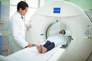
Magnetic resonance imaging (MRI) uses radio waves and magnets to take pictures of organs and structures inside the body by measuring their energy. Like a computed axial tomography (CT) scan, an MRI photographs the organ several times, while a patient lies on a table. A computer joins all the images and creates a 3D image.
Why Use MRI?
Doctors use MRI to help diagnose and monitor pancreatic cancer. Advantages of MRI are:
- Tumors that are not visible on other scans sometimes appear on MRI scans
- People who are allergic to the contrast dye needed for CT scans may prefer an MRI scan since it usually uses a different contrast substance
- MRI scans do not expose patients to radiation
What Is MRCP?
Magnetic resonance cholangiopancreatography (MRCP) is a special type of MRI that uses computer software to image the pancreatic and bile ducts, areas where tumors often form. MRCP is also used to see pancreatic cysts and blockages in the ducts. A MRCP can happen at the same time as an MRI.
Why Use MRCP?

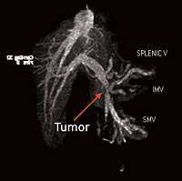
Arrow shows a pancreatic tumor on MRCP scan
MRCP provides a similar picture as an endoscopic retrograde cholangiopancreatography (ERCP), but without the same risks.
Also, MRCP may be used to diagnose other conditions including a type of pancreatic cyst called an intraductal papillary mucinous neoplasm (IPMN).
How Should Patients Prepare for an MRI or MRCP Scan?
Before an MRI or MRCP, patients can eat and take medicines as normal, unless told otherwise. Because these machines use powerful magnets, patients must:
- Remove all clothing, jewelry and medical devices that may have metal
- Wear no makeup, since some products have metals or minerals that may affect the procedure
- Tell the technician if they have any implanted metal or electronic devices, like a heart valve, pacemaker or prosthetic joint
What Happens During an MRI or MRCP Scan?
An MRI or MRCP procedure may take up to an hour. The patient lies on a sliding table inside a narrow tube. The radiation technician is in a room nearby and can communicate with the patient.

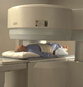
Open MRI
The magnets in an MRI scanner often make loud thumping noises. Patients usually get earplugs or headphones with music to make them more comfortable.
During the test, the patient must lie very still to produce clear images. The doctor and technician can use the computer to rotate the images and view them from different angles.
Patients who are claustrophobic (afraid of closed-in spaces) may need medicine to calm their anxiety before entering a standard MRI machine. Or they can try a different type of MRI scanner called an “open MRI,” where the machine’s sides are open.
What Happens After an MRI or MRCP Scan?
Most MRI and MRCP scans are painless, and there are generally no risks or complications. Patients who may have needed medicine for anxiety should not drive a vehicle and should plan to have someone drive them home.
Before the patient leaves the exam table, the radiation technologist makes sure that the pictures are high quality. After the scan, it may take several days to learn the results from an MRI or MRCP.
How Do MRI and MRCP Compare to Other Imaging Tests?
MRI and MRCP scans are:
- Noninvasive
- Generally low risk
- Radiation-free
MRI images are like those from a CT scan. An MRI scan takes several pictures of the organ, while the patient lies on a table.
A computer combines all the images to create a 3D scan. MRI scans may take longer than CT scans and require the patient to lie motionless in a long cylinder.
MRCP provides a similar picture to ERCP, but without the risks of an invasive procedure.
We’re Here to Help
For more information about how pancreatic cancer is diagnosed, including imaging tests like MRI and MRCP, contact PanCAN Patient Services.
Other Imaging Tests
Computed Tomography or Computerized Axial Tomography (CT or CAT)
Positron Emission Tomography – Computed Tomography (PET-CT)
Endoscopic Retrograde Cholangiopancreatography (ERCP)
Information reviewed by PanCAN’s Scientific and Medical Advisory Board, who are experts in the field from such institutions as University of Pennsylvania, Memorial Sloan-Kettering Cancer Center, Virginia Mason Medical Center and more.
Information provided by the Pancreatic Cancer Action Network, Inc. (“PanCAN”) is not a substitute for medical advice, diagnosis, treatment or other health care services. PanCAN may provide information to you about physicians, products, services, clinical trials or treatments related to pancreatic cancer, but PanCAN does not recommend nor endorse any particular health care resource. In addition, please note any personal information you provide to PanCAN’s staff during telephone and/or email communications may be stored and used to help PanCAN achieve its mission of assisting patients with, and finding cures and treatments for, pancreatic cancer. Stored constituent information may be used to inform PanCAN programs and activities. Information also may be provided in aggregate or limited formats to third parties to guide future pancreatic cancer research and education efforts. PanCAN will not provide personal directly identifying information (such as your name or contact information) to such third parties without your prior written consent unless required or permitted by law to do so. For more information on how we may use your information, you can find our privacy policy on our website at https://www.pancan.org/privacy/.


